Neck And Shoulder Anatomy Diagram. This article describes the anatomy of the head and neck of the human body, including the brain, bones, muscles, blood vessels, nerves, glands, nose, mouth, teeth, tongue, and throat. The clavicle and the manubrium are the ventral borders of the neck area, while the acromion and the. C4 enables you to shrug your shoulders and automatically causes the diaphragm to contract when you are breathing. 8 name the arteries and the. The shoulder is one of the largest and most complex joints in the body. Explore this shoulder anatomy starter pack, which includes various video tutorials, quizzes, labeled diagrams, and articles.
The functions of the levator scapulae include the lateral flexion of the neck (ipsilateral), drawing the scapula superomedially and rotation of the glenoid cavity inferiorly. Despite being a relatively small region, it contains a range of important anatomical features. This post is part of a series called human anatomy furthermore, the trapezius muscle, which from the front appears to connect the shoulder with the neck, is highly individual; Explore this shoulder anatomy starter pack, which includes various video tutorials, quizzes, labeled diagrams, and articles. (see other sets for scapula and upper arm bony anatomy) learn with flashcards, games and more — for free. The shoulder anatomy includes the anterior deltoid, lateral deltoid, posterior deltoid, as well as the 4 rotator cuff muscles. Webmd's shoulder anatomy page provides an image of the parts of the shoulder and describes its function, shoulder problems, and more. Home > blog > anatomy > shoulder anatomy: The neck muscles, including the sternocleidomastoid and the trapezius, are responsible for the gross motor movement in the muscular system of the head and neck.
Anatomy and function neck, regions of the lower face, cervical spine, head joints, cervical organs.
(see other sets for scapula and upper arm bony anatomy) learn with flashcards, games and more — for free. The neck is the area between the skull base and the clavicles. Home > blog > anatomy > shoulder anatomy: For that reason, and because of the dexterity of the shoulder joint itself, the musculature of the shoulder is complex. The sternoclavicular (sc) joint supports the connection of the arms and shoulders to the main skeleton. The 4th cervical spinal nerve. You can see it enclosing the glenohumeral joint and you can see its attachment on the anatomical neck that's the shoulder joint. The shoulder is one of the largest and most complex joints in the body. This webpage presents the anatomical structures found on shoulder mri. The functions of the levator scapulae include the lateral flexion of the neck (ipsilateral), drawing the scapula superomedially and rotation of the glenoid cavity inferiorly.
Interactive anatomical atlas of the head, brain, and neck based on anatomical diagrams and ct and mri medical imaging exams. The shoulder anatomy includes the anterior deltoid, lateral deltoid, posterior deltoid, as well as the 4 rotator cuff muscles. The 4th cervical spinal nerve.

Interactive anatomical atlas of the head, brain, and neck based on anatomical diagrams and ct and mri medical imaging exams.
Related posts of muscle anatomy neck and shoulder. But when i got to thinking of all the things i wanted to tell you about it's very defined on some guys, and hardly present on others. Ct, mri, radiographs, anatomic diagrams and nuclear images. Simple easy notes for quick revision for exams. Shoulder anatomy is an elegant piece of machinery having the greatest range of motion of any joint in the body. 8 name the arteries and the. This diagram here just shows the joint capsule itself. Click on a link to get: Three bones come together at the shoulder joint. Robin smithuis and henk jan van der woude. Clinically, surface anatomy is used to split the neck into anterior and posterior triangles which provide clues as to the location of specific structures. This acts as the bony framework by which the muscles of the chest, upper back and shoulder connect the upper limb to the trunk of the body and control it's movements.the clavicle connects to the sternum via the sternoclavicular joint and to the scapula by. 7 draw labelled diagram showing the relations of shoulder joint.
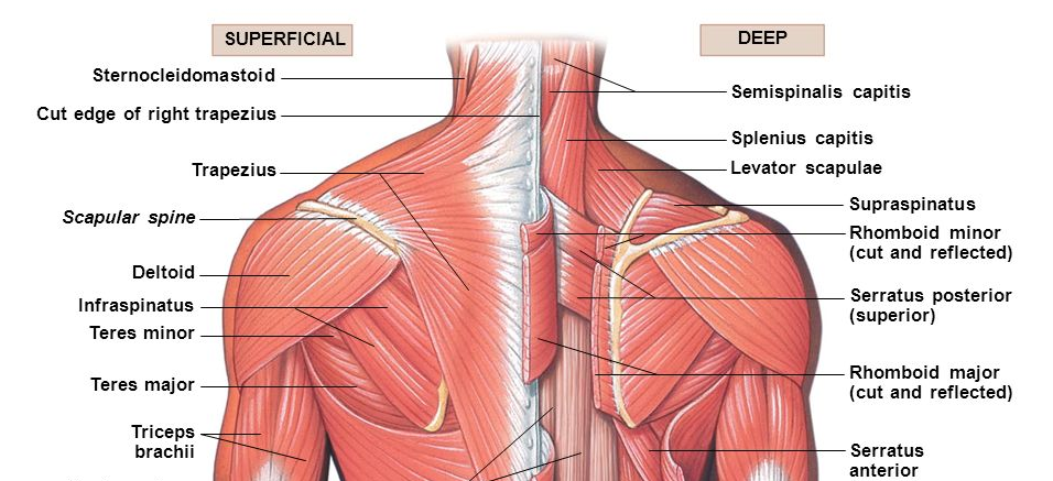
Shoulder muscle anatomy shoulder muscles bicep tendonitis scapula acromioclavicular joint shoulder bones ligaments and tendons shoulder trapezius a large muscle consisting of three parts covering upper back, shoulders, and neck. The 4th cervical spinal nerve. Caudally, the neck is bordered by bony structures of the shoulder girdle and the sternum.

The clavicle and the manubrium are the ventral borders of the neck area, while the acromion and the.
You can see it enclosing the glenohumeral joint and you can see its attachment on the anatomical neck that's the shoulder joint. Click on a link to get: Normal anatomy, variants and checklist. Instead of your doctor simply saying that the patient knee hurts, he or she can say that the shoulder dislocations and surgical neck fractures (breaks) of the humerus can therefore be accompanied by nerve injury. Interactive anatomical atlas of the head, brain, and neck based on anatomical diagrams and ct and mri medical imaging exams. The shoulder joint is formed where the humerus (upper arm bone) fits into the scapula. The head rests on the top part of the vertebral column, with the skull joining at c1. Sechrest, md narrates an animated tutorial on the basic anatomy of the shoulder. Anatomy and function neck, regions of the lower face, cervical spine, head joints, cervical organs. Three bones come together at the shoulder joint. Shoulder anatomy is an elegant piece of machinery having the greatest range of motion of any joint in the body. This diagram here just shows the joint capsule itself.
This diagram here just shows the joint capsule itself shoulder anatomy diagram. Anatomy terms allow us to describe the body and body motions more precisely.

Posting Komentar untuk "Neck And Shoulder Anatomy Diagram"